Dougie, a five-year-old French Bulldog, was recently referred to the Neurology Department at ChesterGates Veterinary Specialists for investigation of acute-onset non-ambulatory Tetraparesis following a fall down the stairs.
Initial treatment with Paracetamol and Gabapentin had not resulted in any improvement, so when Specialist Neurologist Rocio Orlandi arrived, she carried out a thorough neurological examination.
Dougie was alert but unable to stand or walk, with minimal voluntary movement in all four limbs and absent postural responses. Segmental reflexes were normal, and discomfort was noted on cervical spinal palpation. Findings were consistent with a lesion affecting the C1–C5 spinal cord segments, with intervertebral disc extrusion (IVDE) considered the most likely diagnosis.
Dougie was admitted for cage rest and multimodal analgesia, and Rocio scheduled an MRI for the next day. Dougie was monitored closely overnight by the highly trained wards nursing team. However, after a stable night, Dougie suddenly developed hypoventilation, a rare but recognized complication of high cervical cord injury due to impairment of the intercostal muscles.
Rocio, along with the emergency on-call Specialist Anaesthetist Ffion Lloyd rushed Dougie to prep and blood gas analysis confirmed elevated CO₂ levels. Emergency anaesthesia was initiated to support ventilation, and the emergency on-call radiologist was called to carry out an immediate MRI. A scan of the cervical spine revealed a C3–C4 IVDE with associated epidural haemorrhage and significant spinal cord compression.
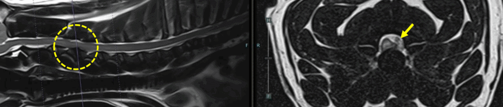
Magnetic resonance imaging (MRI) of the cervical spine revealed a C3-C4 intervertebral disc extrusion alongside epidural haemorrhage with marked left ventrolateral compression of the spinal cord. Generalised intervertebral disc degeneration was also noted.
Dougie was immediately prepped for theatre and surgical decompression via a left-sided hemilaminectomy was performed by Rocio, assisted by theatre technician Sian Holt guiding the intraoperative fluoroscopy to confirm lesion localisation. Ffion monitored Dougie’s anaesthesia closely, ensuring intraoperative stability and analgesia.
After a flawless surgery, Dougie immediately showed a marked improvement in thoracic expansion and respiratory function. Over the following days, he was monitored closely as he regained thoracic and pelvic limb movement until he was finally discharged on day 3 with a tapering course of Prednisone, ongoing analgesia, and a structured rehabilitation plan including restricted exercise and hydrotherapy from week four post-op.
At his one-month recheck, Rocio was extremely pleased to see that Dougie was ambulatory and continuing to improve. She commented: “Dougie’s case was a true team effort. Thanks to our on-site advanced imaging, surgical theatre with fluoroscopy, and seamless collaboration between neurology, anaesthesia, radiology, nursing and intensive care, we were able to respond quickly and give Dougie the best possible outcome.”
ChesterGates Veterinary Specialists is renowned for its expertise in veterinary neurology, offering one of the most advanced neurology and neurosurgery services in the region, and this case highlights the importance of multidisciplinary coordination and timely intervention in the successful management of complex cervical spinal injuries.