News
Cholesteatoma
15th November 2018
A rare cause of ear disease
By Daniella McCready, European Specialist In Small Animal Surgery
Alfie Knowles, a 10-year-old Pug presented to ChesterGates Veterinary Specialists with a six month history of a left head tilt, ataxia and nystagmus.
Neurological examination was consistent with peripheral vestibular disease. Alfie was anaesthetised. Otoscopic examination of the ears and a CT of the head were performed. Otoscopic examination demonstrated a significant amount of white scales within the external ear canal and bulla. The tympanic membrane was ruptured.
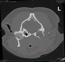
Transverse image of CT of a 10 year old Pug
What are the findings?
There is a moderate amount of soft tissue within the left tympanic bulla. The left bulla is expansile. There is a mixed lytic and proliferative pattern of the left tympanic bulla and lysis of the left temporal bone.
What is the diagnosis?
CT findings were consistent with a left aural cholesteatoma.
Differential diagnoses include aural neoplasia and chronic otitis externa/media.
Cholesteatomas are epidermoid keratin filled cysts characterised by expansile growth and destruction of adjacent tissue esp. bone. These lesions can be mistaken as malignancy. The cause of cholesteatomas is unknown however cholesteatomas have been reported in chronic cases of otitis externa.
What is the treatment and prognosis?
Surgery is recommended with either a lateral or ventral bulla osteotomy and removal of all the keratin debris and epithelium. A ventral bulla osteotomy was performed in this case and the keratin scales/flakes as well as the epithelium from the bulla removed. Histopathology in this case was consistent with a cholesteatoma. A swab from the bulla demonstrated no bacterial growth – confirming a cholesteatoma is a sterile process.
Approximately 50% of cases have concurrent neurological signs such as peripheral vestibular disease and facial nerve paralysis. Unfortunately, neurological signs can persist following surgery and recurrence can occur in 50% of cases. Revision surgery can be performed. Alfie made an unremarkable recovery from surgery. Follow-up examinations demonstrated some neurological improvement with resolution of the nystagmus and ataxia however, a left head tilt remains.
Back to news & events