News
Cases of pulmonic stenosis with different causes – the value of multimodal imaging
8th December 2021
These cases were initially seen by Emily Dutton (RCVS Specialist in Cardiology) and diagnosed with severe pulmonic stenosis. They were referred for angiography-CT to evaluate for the presence of aberrant coronary arteries and subsequent surgery, if suitable. Cardiology specialist Liz Bode, then evaluated both cases prior to performing surgery with the help of our referral clinician in diagnostic imaging, Pablo Menendez.
The first case is an 18 month old male English bulldog.
Pulmonic stenosis is a common congenital heart condition, often observed in brachycephalic breeds, especially English and French bulldogs. In a number of these individuals, we can see abnormal anatomy of the coronary arteries causing, or at least contributing to, stenosis of the pulmonic valve. If an abnormal coronary artery is identified, then balloon valvuloplasty is contraindicated due to the potential for artery rupture and death under anaesthesia. Transthoracic echocardiography is usually supported by angio-CT, with or without transoesophageal echocardiography, when evaluating such breeds for suitability for balloon valvuloplasty. Here at ChesterGates we have all these imaging tools at our disposal.
Bear underwent angio-CT and transoesophageal echocardiography. This confirmed the presence of an aberrant coronary artery and so balloon valvuloplasty was not advised. He was maintained on atenolol, which is prescribed for cases with severe pulmonic stenosis.
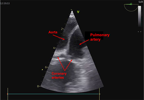
Transoesophageal echo and CT
The second case is an 8 month old male French bulldog. He underwent angio-CT and had normal coronary artery anatomy. Therefore, he was a suitable candidate for balloon valvuloplasty.
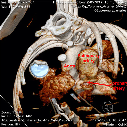
CT image showing normal coronary artery anatomy
Albert’s pressure gradient prior to surgery was 104 mmHg. This means that the right ventricle, which normally operates at a systolic pressure of 20mmHg, was generating a pressure of >120mmHg which is closer to left ventricular pressure. Balloon valvuloplasty was performed under general anaesthesia by Liz and Emily. A balloon catheter was delivered through the right jugular vein into the right ventricle and across the pulmonic valve. The balloon catheter was then inflated several times until loss of the stenosis was seen. Following surgery, the right ventricular pressure had reduced to 58mmHg. A decrease in pressure of around 50% is considered a success.
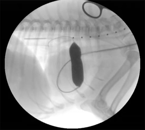
Balloon valvuloplasty
Albert is currently doing very well, and his pulmonic stenosis has decreased from severe to mild-moderate, which means that Albert has an excellent prognosis.
Back to news & events