News
A case of Atlantoaxial Instability – AAI
13th August 2021
This case of AAI was diagnosed by Aran Nagendran (locum neurology specialist) and Ana Martinez (neurology intern) and managed later on by neurology specialist Emili Alcoverro. The surgery was planned and executed by orthopaedic Specialist, Ben Walton, with surgical guides from Fusion Implants.
Chance is a six-month old Maltese dog that was referred to ChesterGates Veterinary Specialists due to generalised incoordination.
Ben Walton explains the case:
“Atlantoaxial instability (AAI) is most often seen in young, small breed dogs. Surgery aims to stabilise the atlas and axis to prevent ongoing spinal cord compression and injury. The significant technical challenge is that the margin for error is very small: holes need to be drilled in small bones very close to the spinal cord and other vital structures, which are not visible to the surgeon. Furthermore, surgical access is restrictive with all of the important structures of the neck in the way.
Custom drilling guides have revolutionised this surgery from the operator’s perspective. CT scans were collected at Chestergates Veterinary Specialists and sent to Fusion Implants. There, James Sage, a skilled biomedical engineer, with clinical guidance from Specialist surgeon, Ben Walton, designed drilling guides based on ideal screw trajectories and bone surface morphology. These were then printed in autoclavable resin and sent back to Chestergates. Neurologist Emili Alcoverro I Balart and Ben performed the surgery together. The screw holes were drilled using the custom guides, and the screws bonded together with polymer. Post-operative CT confirmed perfect alignment and implant positions.
This case perfectly demonstrates the value of a team approach to complex cases. Success here depended on dedicated owners, a rapid diagnosis by the neurology team, seamless cooperation between Fusion and ChesterGates, Specialist anaesthesia input to make the procedures safe, accurate diagnostic imaging, and highly trained and truly caring nursing teams in theatre, wards and beyond.”
Since discharge, Chance’s gait and comfort have been gradually improving and two weeks after surgery, the owners reported that he is back to his normal self.
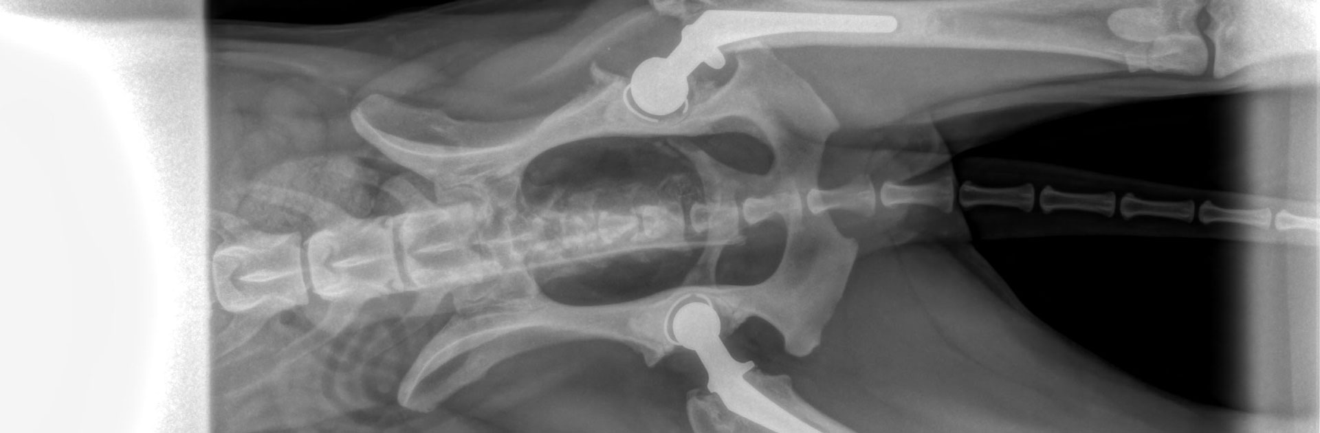
[The bones need to be stabilized with screws, but they are tiny and drilling in the wrong place could be catastrophic! (1a, 1b)]
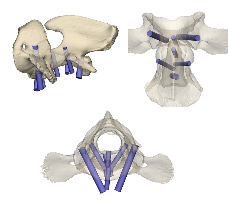
[We plan exactly where we want the screws to go (2a), exactly what size they should be (2b), and exactly what angles they should be at (2c).]
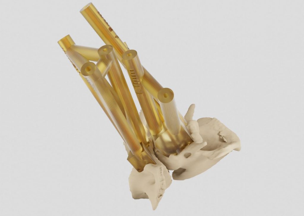
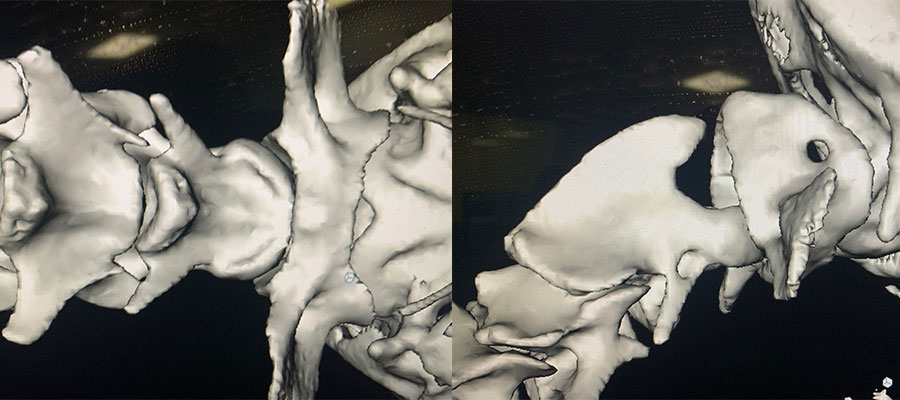
[Then we design and 3D print these drilling guides. They fit the bone surface perfectly and each tunnel is a known length and diameter (3).]
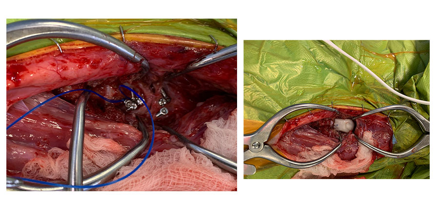
[Next we go to surgery. We drill the holes, insert the screws, and bond them together with polymer.]
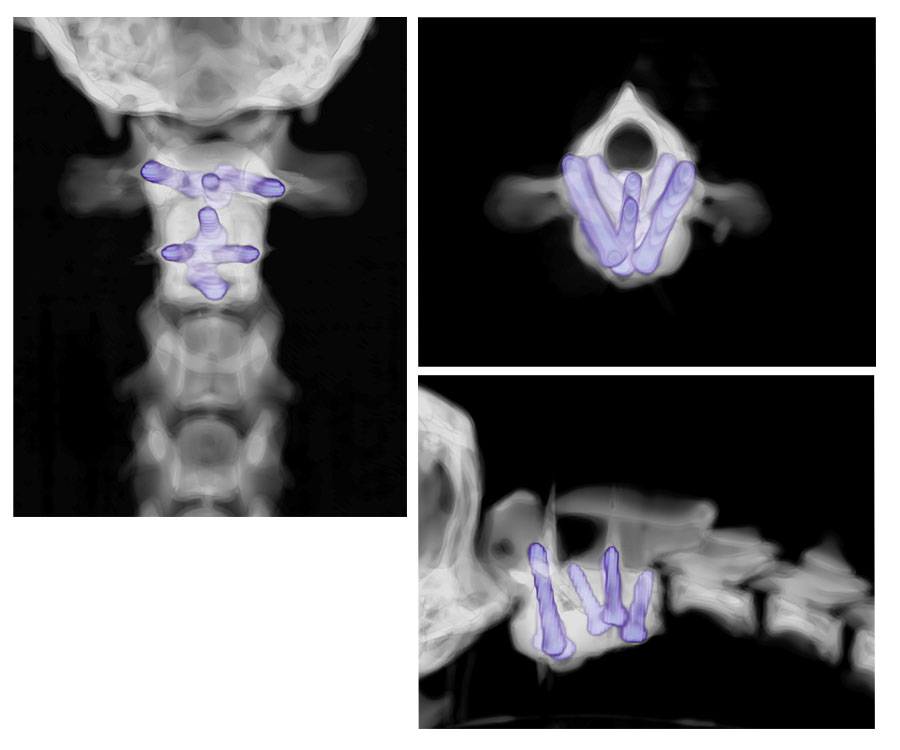
[We then use CT to check the implants are in the correct place (4a), do not impinge on the spinal canal (4b) and that the bones are in the correct position (4c).]
Back to news & events